Exploring the mystery of medial tibial stress syndrome
Is bone overload the main culprit with medial tibial stress syndrome? Does foot pronation play a role in the etiology? Are there any treatments that are consistently effective for this condition? Addressing these questions and much more, this author offers a thorough review of the literature on the common, albeit poorly understood, condition of medial tibial stress syndrome.
Although medial tibial stress syndrome (MTSS) is among the most common musculoskeletal injuries in sport, experts continue to debate the etiology of this injury today. As a result, there is no validation for reliable treatments while myths and misconceptions about this condition continue to abound among athletes, coaches and health-care professionals. Currently, we now consider the injury long-described as “shin splints” a pain syndrome which may result from multiple contributing factors.
The most widely accepted definition of MTSS, proposed by Yates and White, is “pain felt along the middle or distal third of the posteromedial border of the tibia.”1 They further state that aggravation of the pain of MTSS occurs with weight-bearing activity and subsides gradually upon stopping. Clinical examination reveals palpable tenderness along the posteromedial border of the tibia over a length of at least five cm.1
A systematic review of published literature reveals MTSS as the most common running-related musculoskeletal injury with an incidence ranging from 13.6 to 20 percent.2 Verifying this in a subsequent 2018 study, Mulvad and colleagues cited MTSS as the most frequent injury in runners, comprising 16 percent of all injuries.3 This injury is most troublesome among military trainees and has spawned several studies to identify causative factors and remedies for this specific patient population.1,4-7
Pertinent Points Regarding Symptoms And The Differential Diagnosis
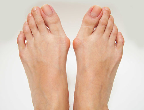
Patients with MTSS describe pain in the distal two-thirds of the tibia along the posteromedial border (see first photo above). Initially, the pain begins with initiation of activity and may actually improve during the exercise session. As the condition progresses, the pain may persist after activity. Persistent pain in the tibia during and after running can also be a sign of a stress fracture. Authorities agree that with tibial stress fractures, pain is more focal while MTSS results in more diffuse pain.7,8 Also, with tibial stress fractures, patients commonly report pain at night, which is reproducible with percussion.8
Erythema and swelling are common with tibial stress fractures but are rare in cases of MTSS.9 Plain radiographs will often be normal with both tibial stress fractures and MTSS.8,10 Therefore, bone scintigraphy and magnetic resonance imaging (MRI) are the best confirmatory studies for tibial stress fractures.10,11
Besides stress fracture, the differential diagnosis of exercise-induced lower leg pain also includes exertional compartment syndrome. Patients with chronic exertional compartment syndrome will complain of neurologic symptoms brought on by exercise-induced elevation of pressure within the deep posterior compartment of the lower leg. These symptoms include cramping, burning or tightness in the leg.12 The symptoms of exertional compartment syndrome arise predictably with the onset of running activity, resolve with cessation of activity and then return with the next training session.13 In contrast, patients with MTSS have pain, which persists for many hours or days after activity.8 Ultimately, exertional compartment syndrome requires diagnosis with intra-compartmental pressure testing.13
Popliteal artery entrapment will cause exercise-induced lower leg pain due to a congenital anomaly along the course of the popliteal artery in the popliteal fossa.14 Claudication occurs from contraction of the gastrocnemius muscle against the abnormal location of the popliteal artery, which is induced by activities such as walking and running.15 To diagnose this condition, one should palpate the pedal pulses bilaterally after exercise with the ankles in full dorsiflexion and knees in full extension as these provocative measures place tension on the gastrocnemius muscle and compress the popliteal artery.16 Reduction of the pedal pulses with these tests is pathognomonic for popliteal artery entrapment syndrome.16
One can diagnose MTSS accurately and reliably using patient history and clinical examination findings.17 Imaging studies are not necessary for the diagnosis of MTSS but may be helpful in distinguishing the condition from a tibial stress fracture.8 Clinicians should be aware that many asymptomatic runners will show signs of periostitis and bone marrow edema of the tibia on MRI, which is a normal adaptation of bone to impact stress.18
Understanding The Potential Etiologies Of MTSS
While MTSS is a pain syndrome, the exact pathology causing the condition remains a subject of debate. Current evidence suggests that MTSS is either a crural fasciopathy, a tibial bone overload injury or combination of the two.8,19
It is interesting to look back at the history of publications on this syndrome as researchers began challenging the simple concept of “shin splints” and eventually proposed a more sophisticated model of impaired bone healing to explain MTSS.20
As early as the 1950s, one can note the term “shin splints” linked to a traction injury of the lower leg musculature.21 The traction theory is based upon a mechanism whereby certain muscles in the lower leg create a periostitis along the tibia at their origin or attachment site.
However, researchers began to challenge the traction theory, which is based upon the anatomy of muscular attachments to the tibia versus the actual site where running athletes most often experience MTSS pain.
The muscle traditionally implicated in causing a shin splint syndrome is the tibialis posterior.22 Saxena and coworkers further promoted this notion in their cadaver study that found that the tibialis posterior originated from the tibia at a location 7.5 cm proximal to the medial malleolus.23 Beck and colleagues subsequently refuted this finding, stating that only the flexor digitorum longus (FDL) and soleus muscles actually originate from the medial aspect of the tibia, most notably in the middle and proximal sections.24
Brown and coworkers also conducted a detailed cadaver study and found that only the FDL and soleus attach to the posteromedial border of the tibia with an absence of attachment of the tibialis posterior at this location.25 Furthermore, they found virtually no soft tissue attachment to the distal one-third of the tibia other than the deep crural fascia.25 Therefore, only the FDL and soleus attach to the posteromedial border of the tibia and these attachments are primarily in the middle and proximal sections of this bone (see illustration above). This does not explain how muscular traction alone can produce MTSS in the typical location at the distal one-third of the posteromedial border of the tibia.
However, the crural fascia, which attaches to the entire length of the posteromedial border of the tibia, may support the traction etiology of MTSS. Several authors propose a mechanism whereby excessive activity of the deep posterior muscle group of the lower leg increases tension in the crural fascia, which then places traction on the periosteum of the tibia.26,27
Can Foot Pronation Be A Contributing Factor With MTSS?
Researchers have suggested that MTSS may result from excessive muscle activity in the lower leg caused by foot pronation.27-29 Specifically, Michael and Holder noted that foot pronation caused excessive elongation strain along the medial origin of the deep posterior muscles of the lower leg during running.30 Richie and coworkers studied athletes running on three floor surfaces and measured electromyographic (EMG) output of the medial tibial musculature.31 This study showed that running on harder surfaces evoked greater eccentric muscle activity along the medial tibia and the magnitude of muscular activity was greater in people with a more pronated foot posture.31
Noting this previous work, Bouché and Johnson conducted a cadaver study to determine if increased loading of the tibialis posterior, flexor digitorum longus and soleus tendons would increase strain in the crural fascia attached to the distal medial crest of the tibia.32 Indeed, this study showed a direct linear relationship between increased tension in the three aforementioned muscles, which then produced strain in the crural fascia at the tibial attachment. Bouché and Johnson proposed that MTSS is actually a “tibial fasciitis,” which results from excessive muscular contraction causing traction on the deep tibial (crural) fascia along the distal-medial tibial crest.32 Other investigators subsequently confirmed this proposed tibial fasciitis mechanism based upon the anatomic location of the deep crural fascia of the leg and its direct correlation to the area of the tibia most commonly associated with MTSS.33,34
Both Richie and Bouché, with their respective coauthors, speculated that foot pronation would cause excessive eccentric loading of the medial tibial musculature.31,32 Several subsequent studies also identified foot pronation as a risk factor for MTSS. Using rearfoot alignment as a measure of pronation, two studies showed excessive eversion of the calcaneus in runners with MTSS in comparison to non-inured runners.29,35 Two other dynamic, three-dimensional kinematic studies of runners showed that various measures of foot pronation were associated with MTSS.36,37 Two studies using the static measures of the Foot Posture Index as well as the standing medial longitudinal arch angle showed a greater risk of MTSS in runners with a pronated foot posture.1,38 Finally, three prospective studies showed that excessive navicular drop, a measure of midfoot pronation, was a risk factor for runners developing MTSS.37,39,40
What Does The Literature Say About Bone Overload Theory?
Another proposed mechanism for developing MTSS is bone overload from repeated bending of the tibia.41-43 Strain from bending normally leads to adaptation of bone via the activation of osteoblasts.44 With repetitive or excessive strains, there may be bone microdamage in which osteoclast activity overrides osteoblast activity, leading to tibial osteopenia.45 This is the same proposed mechanism for the development of a tibial stress fracture.45
In the tibia, the site of greatest bending stress during running is at the junction of the middle and distal thirds of the bone.46 Studies have shown that people with MTSS have reduced bone mineral densities at this location, which returned to normal after symptoms disappeared.43,47 In a 2019 study, Winters and team evaluated biopsies taken from the painful sites of the tibia in patients with MTSS and noted microcracks without evidence of a repair process.48 They suggested that MTSS may represent a process in which an inhibition or dysfunction in the cell signaling system prevents a remodeling response in the tibia. Winters and colleagues also speculated that the bony microcracks in MTSS were the result of torsion and compression forces rather than tensile forces from muscle or fascial traction.
In their critical review of MTSS, Moen and colleagues cite four important findings that support the bony overload theory for the pathophysiology of MTSS.8 The findings were as follows:
1. Three-phase bone scans are abnormal in the final phase in patients with MTSS, indicating involvement of bone and periosteum;
2. High resolution computed tomography (CT) scans of the tibial cortex demonstrate osteopenia in patients with MTSS, indicating bone remodeling;
3. MRI images of patients with MTSS show bone marrow edema of the tibia as well as an abnormal signal along the periosteum; and
4. There is reduced bone mineral density in patients with MTSS in comparison to healthy controls. When symptoms improve, the bone density returns to normal values.42,43,47,49-52
What Other Studies Have Revealed About Possible Risk Factors For MTSS
Two recent systematic reviews looking at risk factors for MTSS showed similar findings.53,54 One review focused on studies of runners while the other review analyzed studies of physically active individuals, including military personnel. These systematic reviews found greater body mass index (BMI), navicular drop, ankle joint plantarflexion and hip external rotation in patients with MTSS than in non-injured controls.53,54 One can only speculate how these four risk factors might support theories for either the fascial traction or tibial bending mechanisms for MTSS etiology.
Certainly, BMI could account for greater tibial overload during running. Greater external hip rotation may cause increased torque on the tibia during running.55 Increased ankle joint plantarflexion, a risk factor for patellar tendinopathy, may be an indication of greater push-off effort by the runner.56 This exaggerated ankle joint plantarflexion during running might also be related to compensation for midfoot instability, which one can assess by measuring navicular drop.57
Navicular drop may be the most important observation when evaluating risk factors and the pathomechanics of MTSS.37,39,40 Navicular drop is the measurement of the height of the navicular from the supportive surface when an individual is seated with the partial weightbearing foot in subtalar neutral in comparison to the same measurement when the individual is standing unilaterally on the same foot in relaxed stance (see last two photos above).58 Navicular drop is a measure of midfoot pronation and not necessarily rearfoot pronation.59
Excessive rearfoot eversion did not appear to be a risk factor for MTSS according to a meta-analysis of five specific studies.54 However, another systematic review of five other studies, without meta-analysis, confirmed that rearfoot eversion or pronation was a consistent finding in athletes with MTSS in comparison to those without MTSS.60 Interestingly, this same review found that excessive internal tibial rotation was not associated with MTSS. In a recent 2020 study, Langley and colleagues showed an interesting finding in runners with MTSS who demonstrate a loss of coupling between rearfoot eversion and tibial internal rotation in comparison to healthy controls.61 These study authors speculate that the conversion of rearfoot eversion to inversion during midstance, without a reversal of lower leg internal rotation, could create torque in the tibia and cause excessive bone stress.
Putting Together The Evidence: What Causes MTSS?
Although histologic evidence and imaging studies provide strong support for the bone bending theory of etiology of MTSS, one cannot ignore the evidence for the role of muscular traction in contributing to this injury. Furthermore, of all the risk factors, the presence of excessive midfoot mobility as measured with navicular drop is a critical factor to consider in patients with MTSS.37,39,40 Several studies speculate that excessive hindfoot and midfoot pronation recruit activation of the tibialis posterior, flexor digitorum longus and soleus muscles to compensate for a loss of foot stability during key phases of the running gait cycle.29-31,62
Based upon the current evidence, most authorities speculate that MTSS is a result of a combination of accumulated bone stress without repair, possibly exacerbated by excessive traction on the deep crural fascia of the lower leg, created by excessive eccentric muscle loading.8,32,62 Muscles create more bone stress than impact force, especially when there is application of muscular force non-axially and to the periphery of long bones as one would see in the attachment of muscles and the crural fascia to the posteromedial border of the tibia.63 Inexplicably, few investigators have sought to develop treatment interventions that specifically address the current knowledge of the pathomechanics of MTSS.
Current Concepts In The Prevention And Treatment Of MTSS
In 2013, Winters and colleagues published a systematic review of 11 randomized clinical trials studying the effectiveness of various conservative interventions for the treatment of MTSS.64 After scrutinizing studies of low-energy laser treatment, stretching and strengthening exercises, sports compression stockings, leg braces and pulsed electromagnetic fields, the authors concluded that none of the treatments showed positive effects. Studies of iontophoresis, phonophoresis, ice massage, ultrasound, periosteal pecking and extracorporeal shockwave therapy showed moderate effectiveness based upon Level 3 to 4 evidence.64 Of all the treatment options studied, Winters and coworkers concluded that extracorporeal shockwave had the most promise.64
What is surprising is the dearth of studies implementing custom or prefabricated foot orthoses as a preventive measure or as a treatment intervention for MTSS. Given the volume of evidence that excessive rearfoot pronation and midfoot pronation are both significant risk factors for MTSS, it would seem logical that addressing those postural abnormalities with foot orthoses would be a primary treatment approach.37,39,40,60,61 However, only two studies have been conducted using this approach and both showed positive effects with cushioning inserts as well as prefabricated arch supporting orthotics in preventing MTSS in military personnel.65,66 However, the treatment effects of custom foot orthoses for MTSS have not been studied in any randomized controlled trials to date.
Despite the lack of evidence for reliable treatments of MTSS, Winters believes that observational studies strongly suggest that rehabilitation and effective load management should be key strategies in treating this common running pain syndrome.67 At the same time, a different study by Winters and colleagues warns that recovery from MTSS can be a long process.68 They point out that an athlete with greater than three months of shin pain may not have symptoms resolve for up to nine months despite significant modification of activity and loading. To monitor the recovery process, they developed a “MTSS score” which has good validity, reliability and responsiveness.68 This tool allows for assessment of treatment effects based upon patient evaluation rather than relying upon conclusions made by the clinician. Still, the implementation of a gradual loading program for MTSS requires skill and careful monitoring by an experienced rehabilitation specialist, who can integrate strengthening with controlled impact exercise regimens.69,70
In Summary
Despite MTSS being a common running injury, there are currently no effective, reliable treatments. This is a reflection of the fact that the etiology and pathophysiology of MTSS remain obscure and continue to be the subject of ongoing debate. Current evidence supports a theory that MTSS represents a failure of bone healing and remodeling in the tibia that should otherwise occur in response to the accumulated stresses induced during running. Newer strategies in rehabilitative medicine that focus on graded loading exposure appear to offer the best promise for recovery from this common pain syndrome.
REFERENCES